The cochlea is a complex coiled structure. We are objective in our recommendation.
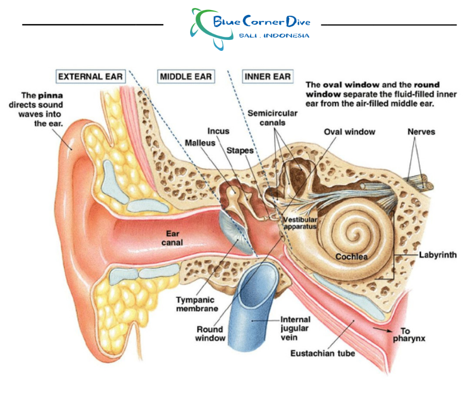 What Happens To My Ears When I Scuba Dive Blue Corner Dive Freediving Conservation
The main difference between inner and outer hair cells is that the inner hair cells convert sound vibrations from the fluid in the cochlea into electrical signals that are then transmitted via the auditory nerve to the brain whereas the outer hair cells amplify low-level sounds that enter into the fluids of the cochlea mechanically.
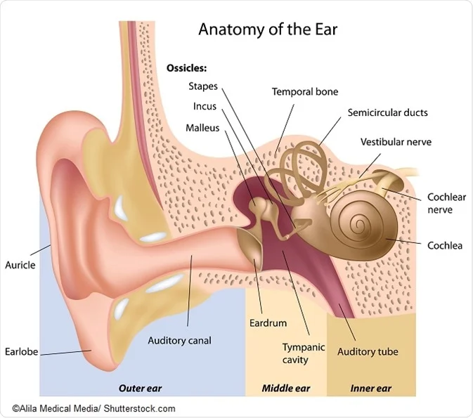
What does the cochlea do. These cochlear hair cells convert audio signals into nerve impulses that are then sent to the brain for processing. The cochlea is a spiral tube that is coiled two and one-half turns around a hollow central pillar the modiolus. The cochlea auditory inner ear transforms the sound in neural message The function of the cochlea is to transform the vibrations of the cochlear liquids and associated structures into a neural signal.
It is located within the inner ear and is often described as hollow and snail- or spiral-shaped. Ad Price is subjective. Sound is collected by the outer ear travels down the air-filled ear canal and hits the eardrum.
We are objective in our recommendation. The brain then translates the impulses into sounds that we know and understand. Structure of the cochlea The cochlea contains the sensory organ of hearing.
The inner earstructure called the cochlea is a snail-shell like structure divided into three fluid-filled parts. This is what produces the sensation of sound. While the cochlea is technically a bone it plays a vital role in the function of hearing rather than simply being another component of the skeletal system.
People with cochlea damage need a greater level of sound to hear well but when sounds get louder they become just as intolerant of loud sounds as people with no hearing impairment. However these cochlear hairs can become damaged for any number of reasons. The cochlea is a coiled structure that resembles a snail shell cochlea comes from the Greek kochlos which means snail.
It is a small--yet complex--structure about the size of a pea that consists of three canals that run parallel to one another. The cochlea lies just behind a membrane called the oval window which separates the middle ear bones from the inner ear. Cochlea are tiny hair cells that vibrate when exposed to sound waves.
Inside the inner ear aka. The other half of the inner ear labyrinth is the vestibular system which controls balance. JACOPIN BSIP Getty Images.
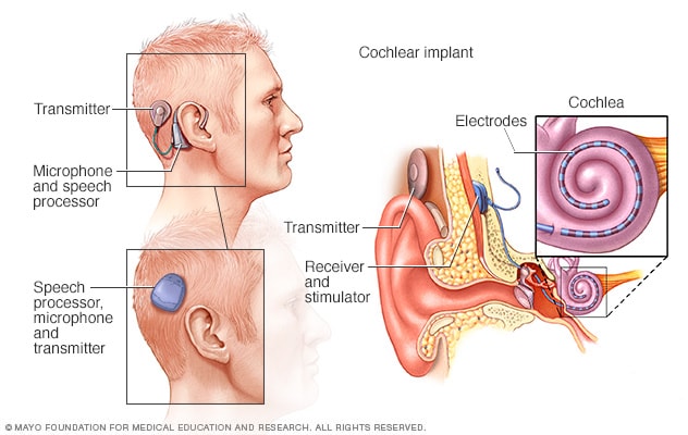
The scala vestibuli scala media and scala tympani. A cochlear implant is an implanted electronic hearing device designed to produce useful hearing sensations to a person with severe to profound nerve deafness by electrically stimulating nerves. In the cochlea sound waves are transformed into electrical impulses which are sent on to the brain.
There are several components of the structure and these combined with the cochlea itself make up the half of the inner ear structure that controls hearing. As seen in audiometry this leaves the patient with a reduced dynamic range. This occurs at the organ of Corti which is located all along the cochlea.
Inner and outer hair cells are the receptive cells found in. Ad Price is subjective. The cochlea is a small snail-shaped bone structure in the inner ear.
Two are canalsfor the transmission of pressure and in the third is the sensitive organ of Corti which detects pressure impulses and responds with electrical impulses which travel along the. It is found within the inner ear. It bears a striking resemblance to the shell of a snail and in fact takes its name from the Greek word for this object.
What is the cochlea and what is the function of the cochlea. This occurs at the organ of Corti a structure consisting of tiny hairs throughout the cochlea that vibrate and send electrical signals through the nervous system. The cochlea contains the organ of Corti with hair cells that detect sound waves in fluid that fills the inner ear organs.
The cochlea is the auditory center of the inner ear a fluid-filled organ that translates the vibrations of auditory sound into impulses the brain can understand. It consists of a long membrane known as the basilar membrane which is tuned in such a way that high tones vibrate the region near the base and low tones vibrate the region near the apex.
Ear Middle Ear Cochlea Cochlea
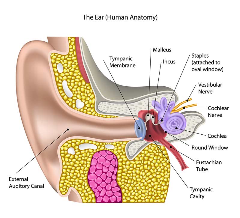
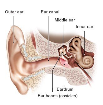 How Does The Ear Work Informedhealth Org
 What Is The Cochlea Definition Function Location Video Lesson Transcript Study Com
 Cochlear Implant Cost Pros Cons Risks How It Works
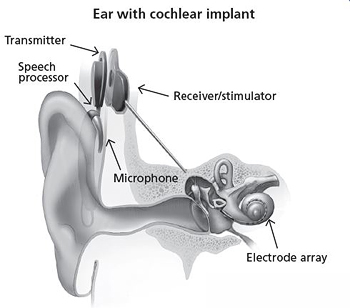 What Are Cochlear Implants For Hearing Nidcd
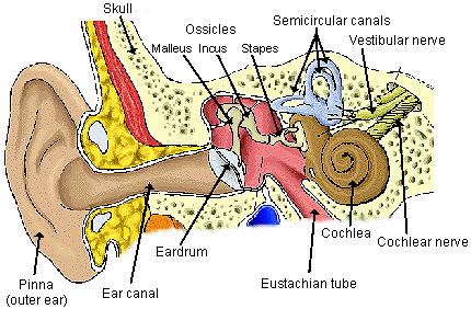 Neuroscience For Kids Audition
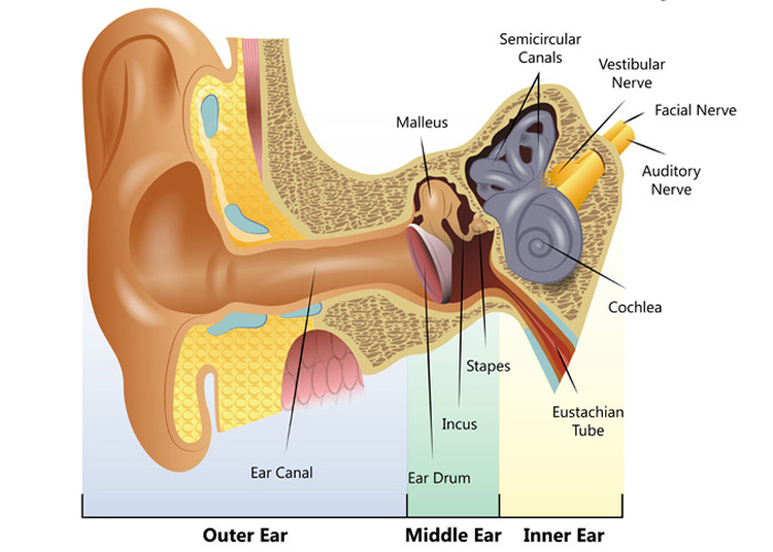 Understanding How The Ear Works Hearing Link



No comments:
Post a Comment
Note: Only a member of this blog may post a comment.