There are no optical aids that could enable the human eye to see something that small. It requires an electron microscope often a scanning electron microscope and yes they are expensive.
 How To Catch The Flu Under The Lens
Light microscopes are the most.

What microscope is used to see viruses. Viruses cannot be seen with optical magnifying microscopes which go up to 1000 times magnification and can be imaged only by electron microscopes. The energy source used in the electron microscope is a beam of electrons. Viruses cant be seen with an optical microscope.
The viral particles are coloured yellow as it emerges from the surface of a cell which is coloured blue and pink. Viruses and some large molecules can be. Apr 27 2012 sophisticated microscopes can see a virus have greater capability of optical microscopy flouresens which can be used to see the inside of.
If the conventional microscope uses light waves the electron microscope instead exposes the sample in a beam of electrons. Here are the most common microscopy techniques used to observe viruses. A fast low-cost technique to see and count viruses or proteins from a sample in real time without any chemicals or dyes could underpin a new class of devices for rapid diagnostics and viral.
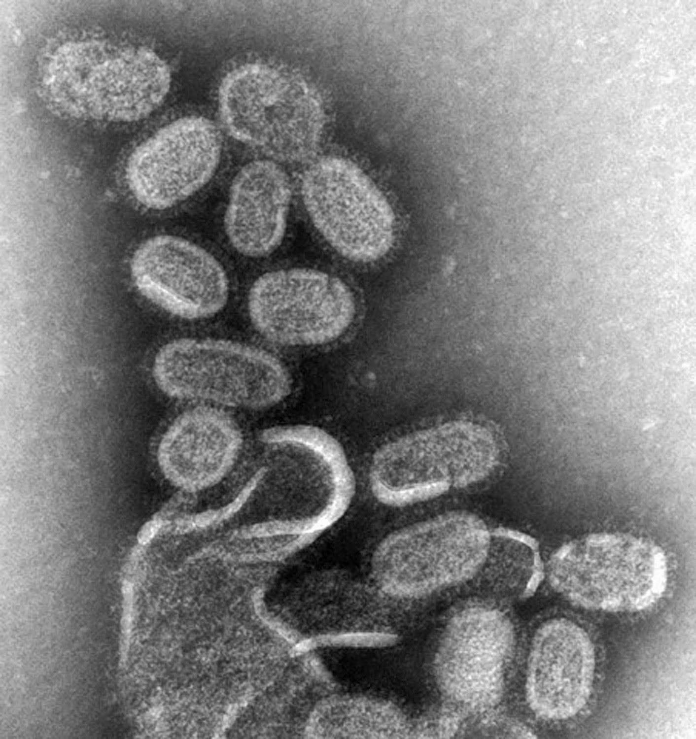
The light microscopes are the types of microscopes used in anatomy and physiology to observe small animals plants metal samples and microorganisms like bacteria in detail. The image was captured and color-enhanced at the NIAID Integrated Research Facility IRF in Fort Detrick Maryland. Since electrons dont travel far in air the chamber where the specimen goes must maintain vacuum conditions.
The image above was captured with a. Potentially one could use a scanning tunnel electron microscope STEM but this a not a good way to screen for viruses because there are not that many such microscopes around because sample preparation takes too long and because it is not really a quantitative readout. The light microscope can magnify a specimen about 1500x and used in many areas of biology anatomy and physiology.
A transmission electron microscope was used to capture SARS-CoV-2 virus particles isolated from a patient. What youre seeing above is a scanning electron microscope image in false colour showing the COVID-19 virus from a patient in the US. Only optical fluoresce microscopes can see inside a virus and then only indirectly using dye which cannot actually penetrate a virus.
The key to the effectiveness of the electron microscope is already in its namethe electrons. Click through the slideshow above to see 50 striking electron micrographs of some of the worlds most dangerous and deadly disease-causing viruses. Microscopes are frequently used equipment in biological sciences that allow us to see organisms and cellular structures that would otherwise be far too small to see.
One of the challenges. Besides how have microscopes been useful to us. Electron microscopes can be used to see viruses viruses are smaller than bacteria so you cant see them using optical microscopes Click to see full answer.
Microscopes allow humans to see cells that are too tiny to see with the naked eye. There are various types of electron microscopy used to image virus particles including immunoelectron microscopy electron tomography transmission electron microscopy and cryo-electron microscopy. Since the beam has an exceptionally short wavelength it strikes most objects in its path and increases the resolution of the microscope significantly.
Mar 1 2011 worlds most powerful optical microscope could view live viruses electron microscopes which use a focused beam of electrons instead of small fry - suggestively.
