Showing posts with label skin. Show all posts

Saturday, February 27, 2021

Black Mole On Skin

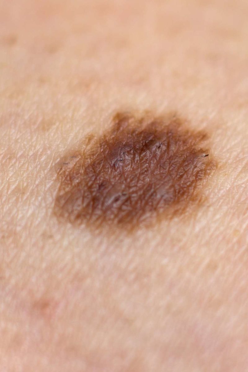
It normally is a flat mole with uneven edges and an asymmetrical shape. Atypical moles are most common in melanoma patients.

Moles Picture Image On Medicinenet Com

The borders of atypical.

Black mole on skin. Benign moles are usually brown tan pink or black especially on dark-colored skin. The risk increases with an increase in the number of these moles. The black bump is a melanoma that is about 3 millimeters wide about 18 inch.

They are common as we get older most often seen as numerous little red dots from 1mm to around 4mm in size but can be solitary and larger. Haemangiomas are essentially blind-ended pouches of blood vessels. Clogged pores that turn black when the air oxidizes the sebum oil the skin produces plug in the pore.

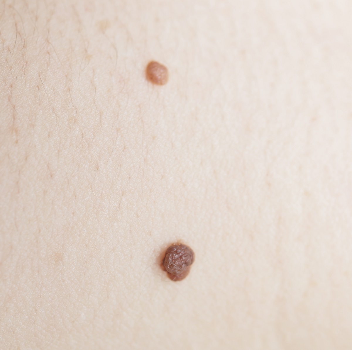
According to the American Academy of Dermatology the most common types of moles are skin tags raised moles and flat moles. This type of black mole is likely to show itself up as a new growth on the skin when one is past the age of 20 years. The melanoma is about 15 millimeters wide or about as wide as a tube of lip balm.

It can be either flat or raised. Very dark skin growths that are usually harmless like skin tags or normal moles but can sometimes be the start of skin cancer. A mole or nevus is a dark spot on our skin comprised of skin cells that have grown in a group rather than individually.

Atypical moles also known as dysplastic nevi are larger than a pencil eraser and shaped irregularly. A melanoma with three partsa dark brown or black area on the left a red bump on the right and an area that is lighter than the skin at the top. Moles are generally less than 6 millimeters about ¼ inch across about the width of a pencil eraser.

This blog is not a substitute for a medical opinion-if you are worried about a changing or funny looking mole or spot get it checked by a doctor with suitable skills. It can be round or oval. Melanoma is a kind of skin cancer that could manifest itself in the form of a black mole on back.

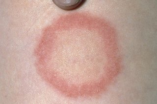
This is a textbook haemangioma a completely harmless skin lesion. Most moles appear by age 30. These moles are usually uneven in color with a dark brown center.

While the healthy mole left has a fairly consistent borderat least as far as mole borders gothe mole on the right has no real perimeter Dr. Dermoscopy with training can help with diagnosis. New moles commonly appear at times when your hormone levels change such as during pregnancy.

They can be any colour that blood is-red blue or black if thrombosed. Moles usually emerge in childhood and adolescence and change in size and color as you grow. Dysplastic nevi also known as atypical moles are another type that is likely to develop into melanoma.

Melanoma skin cancer can be fatal unless caught early but most skin lesions are harmless. Ad Emuaid Gave Me My Life Back I Am So Thankful For This Amazing Product. Dark or black marks on the skin that are due to hyperpigmentation caused by melanin in the skin.

Sun exposure in childhood causes an increase in the number of moles. Ad Emuaid Gave Me My Life Back I Am So Thankful For This Amazing Product. These cells are called melanocytes and are responsible for producing melanin the pigment color in our skin.

A normal mole is usually an evenly colored brown tan or black spot on the skin. They are circular or oval and are usually small commonly between 13 mm though some can be larger than the size of a typical pencil eraser 5 mm. Moles may be flat or raised.

People who have them can develop either single or multiple melanoma. Moles are common small flesh-colored tan brown or black spots on the skin. Some moles can be present at birth but most appear during childhood or young adulthood.

Sunday, September 27, 2020

Skin Lesion Guide

Benign lesions can be classified by their cellular origin. Nevertheless it is useful to know how the common skin lesions look like to be able to recognise them.

Ems Solutions International Marca Registrada Skin Lesion Guide Atlas

Visual Guide to Skin Lesions in Mast Cell Diseases.

Skin lesion guide. Freckles and flat moles are types of macules. Symmetry in shape colour and structure. Ad Emuaid Gave Me My Life Back I Am So Thankful For This Amazing Product.

Superficial elevated solid flat topped. Note if the colour appears consistent throughout the lesion. Assess the diameter of the skin lesion.

Benign skin lesions can present themselves in many different ways from freckles and moles to skin tags and age spots. Our skin guide is a comprehensive guide for 100 skin diseases. The presence of multiple colours within a single skin lesion is suggestive of malignancy.

A doctor can look at each case to determine the right treatment. Vesicle containing pus inflammatory cells Plaque. Measure the size of the skin lesion and ask the patient if it has been growing in size.

A few skin lesions resemble malignancies. 01 The structure and function of the skin 3 02 Taking a history 11 03 Examining the skin and describing lesions 17 04 Common skin conditions in adults 29 Acne31 Cellulitis35 Psoriasis37 Shingles herpes zoster 40 Skin cancers 42 Vasculitis49 05 Common. Macules are another skin lesion that appears like small spots and brown.

Melanocytic keratinocytic vascular fibrous fat. History Questions to ask. Stable or slowly evolving.

Knowing the similarities and. While cutaneous mastocytosis can include maculopapular cutaneous mastocytosis MPCM formerly known as urticaria pigmentosa UP. Absence of spontaneous bleeding bleeding or ulceration may be due to recent injury.

Common Nonneoplastic Skin Lesions Kelly Liesse MS3. Skin Lesion Reference Guide Bulla Circumscribed collection of free fluid 1 cm Macule Circular flat discoloration. Skin Lesion Guide - Atlas Diagnosis Dermatology Skin Lesions Terminology Nomenclature Atlas Key Basics.

The following pages are a photo journal of examples of how mast cell diseases can present. Circular elevated solid lesion 1cm. Most people have them and an overwhelming majority of them are benign.

Other types of primary skin lesions are pustules pus-filled bumps rashes etc. Common skin conditions explained. Progressively enlarging skin lesions particularly those over 6mm in diameter are suggestive of malignancy.

Skin lesions are extremely common. The majority of skin lesions are benign but when a new lesion or mark appears on our skin it can be difficult to tell whether it is dangerous. Secondary skin lesions evolve from primary skin lesions.

Assess the colour of the skin lesion. A majority of the pictures are of skin manifestations of mastocytosis. The treatment may include medication surgery or home care in the case of harmless skin lesions that only need to.

Onset Pattern Skin symptoms itching warmth swelling peeling etc Systemic symptoms fever chills cough NV etc Related factors Environmental or occupational exposures Other medical or comorbid conditions Prescription OTC and illicit drug use Sick contacts. Whilst many people are not fazed by their lumps and bumps others may feel self-conscious by their appearance and therefore seek removal treatment. Your Dermatology Pocket Guide.

Lesions that are growing spreading or pigmented or those that occur on exposed areas of skin are of particular concern. Ad Emuaid Gave Me My Life Back I Am So Thankful For This Amazing Product. A Partial Guide to.

Many pictures of skin diseases that you can compare with actual skin concerns and then read Get checked. A nodule is simply the solid skin formation on your skins surface. Treatment of skin lesions can vary based on the form body and other characteristics of each lesion.

The features in common for benign skin lesions include. Black eschars are collections of dead skin that can arise from infarction which may be caused by infection eg anthrax angioinvasive fungi including Rhizopus meningococcemia calciphylaxis arterial insufficiency or. In this tutorial we aim to provide a simple step-by-step guide to anyone who wants to work on the problem of skin lesion classification regardless of their level or.

If you have any suspicions about a mark mole or lesion you should ask your doctor to check it. Black skin lesions may be melanocytic including nevi and melanoma.

Tuesday, March 31, 2020

What Does Lupus Look Like On The Skin

It is often difficult to distinguish whether a face rash is the result of sunburn rosacea or SLE. A lupus rash can appear in the following ways.

Chronic Cutaneous Lupus Erythematosus Discoid Lupus Erythematosus Download Scientific Diagram

Women are much more likely to get SLE.

What does lupus look like on the skin. Patchy redness that can cause scarring Photosensitivity. Skin disease in lupus can cause rashes or sores lesions most of which will appear on sun-exposed areas such as the face ears neck arms and legs. 10 While ACL lesions can sometimes discolor the skin they generally do not scar.

Discoid lupus lesions which are thick and disk-shaped. This type of lupus can affect many organs including the skin kidneys and joints. Ad Read Janes Story About One Simple Trick To End Lichen Sclerosis Fast.

Over time lupus dermatitis photos below page makes their skin noticeably thickened. A common skin condition rosacea may appear as red skin on the face with telangiectases or even pus-filled bumps that resemble acne. Most types are more common in women and often appear between 20.

A scaly butterfly-shaped rash that covers both your cheeks and the bridge of your nose This rash will not leave any scarring in its wake but you may notice some skin discoloration such as dark or light-colored areas. The sun typically triggers the rash or lesions and affects areas such as the face ears neck arms and legs. Non-healing erosion can clearly explain what dermatitis on the hands looks like without treatment.

The disease begins with the usual redness of the skin its reaction to the irritant. Skin rash in reaction to sunlight exposure Mucus membrane ulcers. Ulceration of the lining of.

Skin can be sensitive and easily irritated. They can appear on your arms shoulders neck or any. Discoid lupus occurs in people with chronic cutaneous lupus CCLE.

Discoid skin rash. Several types of lupus affect the skin. People suffering from discoid lupus have a version of the disease that is limited to the skin.

Cutaneous lupus erythematosus is the umbrella term for lupus that primarily affects the skin. Some individuals have or will develop a type of skin disease called cutaneous lupus erythematosus. It gets its name from the coin-shaped lesions it.

A dermatologist a physician who specializes in caring for the skin should treat lupus skin. This type of condition can cause rashes or sores lesions. Contact dermatitis rash picture 2 is.

What Does a Lupus Rash Look Like. Cutaneous affects the skin lupus. It often begins between 15 and 45 years of age.

It is characterized by a rash that appears on the face neck and scalp but it does not affect the internal organs. They may be red and scaly but they do not cause pain or. They often appear on the scalp or face and can cause permanent scarring.

Lupus that affects the skin CLE comes in different forms and causes different types of rashes. Discoid lupus discoid lupus erythematosus is a chronic autoimmune disease affecting the skin. Less than 15 of patients with discoid lupus progress into the systemic form of the disease.

Acute cutaneous lupus ACL is characterized by flattened areas of red skin that appear on the face in a distinctive butterfly-shaped pattern also known as a malar rash. Flares are not. Smoking may increase the risk of developing SLE.

Red ring-shaped lesions that do not itch or scar. Ad Read Janes Story About One Simple Trick To End Lichen Sclerosis Fast. When patients who have Lupus SLE or even discoid along with additional autoimmune diseases experience flare-ups these vary drastically from one individual to another.

The photosensitive lesions may also develop on the arms legs and trunk. Systemic lupus erythematous SLE.

Saturday, August 17, 2019

How To Get Rid Of Skin Lesions

Use a daily lotion that prevents the buildup of dead skin and help soften your skin like a glycolic 10 alpha hydroxy beta hydroxy or Amlactin. The treatment goes after identifying the skin lesion type.

How Do You Get Rid Of Skin Lesions Westgate Family Physicians Westgate Family Physicians

It exfoliates the top layer of the skin with tiny crystals using a handheld device.

How to get rid of skin lesions. Do you feel little bumps around your neck shoulders face or eyelids. This treatment is limited for cancers on the skins surface not deeper ones. If you are wondering how to get rid of white spots on skin from sun there are a number of available.

Apply physical treatment. As a result it improves the collagen and elastin within the skin removing white spots. Primary Skin Lesions Treatment.

Apply physical treatment. A Verified Doctor answered A US doctor answered Learn more. This will help you to get rid of the skin condition.

It also makes the skin appear youthful and rejuvenated. They start dark like a burn that part falls off leaving a light the mid and drk around. The Triple Action Cleanser and the Salux Cloth exfoliate skin in the shower using a combination of salicylic acid glycolic acid exfoliating beads and a little friction.

Always clean the lesion to. In order to improve how the tea tree oil works on the keratosis you need to apply it for up to two or three times in a day. The healing of some lesions can be sped up by applying a bandage.

Always clean the lesion to. Periodically i get sm 2mm or less skin lesions. Photodynamic therapy PDT is especially effective as a one and done treatment to destroy lesions without harming healthy tissue.

Make sure to use a strong exfoliating product but do not over scrub. Its sprayed out of a can and is approximately -320. In order to be on the safe side you should ensure that you continue with the application for up to a week after the keratosis had been removed from your skin.

Skin becomes soft hydrated and smooth. After analyzing all this along with the patients overall health the treatment actually starts. In order to treat any kind of skin lesion the basic thing required is to check is the type of skin lesion which can be either primary or secondary skin lesions.

Chemotherapy involves using cancer-killing drugs in ointments or creams that are applied directly to the skin lesion. Small Skin-Tag Removal If the skin tag is very small the first thing Geraghty does is spray the bump with liquid nitrogen a nontoxic substance. Others such as cold sores will heal faster if you regularly apply ice.

Others such as cold sores will heal faster if you regularly apply ice. You might have panicked and confused them with something dangerous but relax. Apply the Glycolic Acid Body Lotion after toweling dry and your barnacles will slowly start to soften.

The healing of some lesions can be sped up by applying a bandage. For widespread AKs located on the face and scalp a light-sensitizing topical agent is applied for a period of time followed by exposure to blue or red light to kill cancer cells.

Thursday, September 20, 2018

Ring Rash On Skin

Ringworm is described as a red scaly and itchy patch that has a ring shape. Its caused by the fungus dermatophytosis or tinea.

Ringworm Nhs

This occurs when your skin comes into contact with an irritant that causes a reaction.

Ring rash on skin. The Ring-Shaped Rash One of the most distinctive signs of tinea corporis is the appearance of an itchy red circular rash in the shape of a ring. The fungi responsible for ringworm thrive in hot moist environments such as the tropics or more. On lighter skin a ringworm rash is red or pink and on darker skin it is silver or brown.

Ringworm is a common type that mostly affects the skin hair and nails. Ringworm is a misnomer since a fungus not a. Ringworm also known as dermatophytosis dermatophyte infection or tinea is a fungal infection of the skin.

Ring rash is a common skin condition that typically requires minimal at-home treatments. It causes a scaly pink patch about 210 centimeters cm in diameter to. Signs of a skin rash or infection develop in someone with a chronic illness or weakened immune system.

Finding one of these is a helpful way to distinguish this rash from other common skin rashes such as eczema. Because it often looks like a ring of small skin-colored pink or red bumps granuloma annulare might be mistakenly identified as ringworm. Feet with ringworm infection athletes foot Itching burning and stinging on your soles and between your toes Dry scaly skin that usually begins between the toes and can.

This rash may resemble a target or a bullseye and it usually has raised edges. The rash is getting worse and treatment for psoriasis or ringworm does not help. Its not dangerous and can be treated with antifungal medication like Fugacil.

Pityriasis rosea is a common mild skin rash that usually lasts for 68 weeks. Read full article on ringworm. Sometimes ring rash is caused by contact dermatitis.

Ringworm is a common fungal infection and it usually forms a circular or oval-shaped rash. The diameter of the rings can be up to 2. Circular-shaped scaly rashes with raised border.

Skin in the middle of the ring appears clear and healthy and the edges of the ring may spread outward. The skin around the rash may be dry or scaly and the rash. A ringworm infection on the skin is indicated by roundish flat patches that have a raised scaly border.

Jewelry containing nickel or gold can cause allergic. However it is important to find the underlying cause which could be an allergic reaction to metals or. Ringworm is a common fungal skin infection otherwise known as tinea.

Ad Emuaid Gave Me My Life Back I Am So Thankful For This Amazing Product. Ad Emuaid Gave Me My Life Back I Am So Thankful For This Amazing Product. Ringworm most commonly affects the skin on the body tinea corporis the scalp tinea capitis the feet tinea pedis or.

What Are The Signs Of Meningitis

Spots or a rash. Loss of limbs amputation of affected limbs is sometimes necessary. Meningitis Symptoms Signs Of Meningitis Meningitis No...